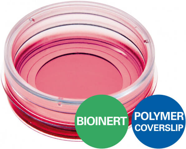
µ-Dish Bioinert

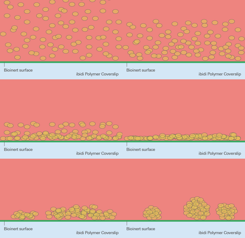
Bioinert 系列產品底部經特殊材質「Polyol hydrogel」處理,比一般 ULA (ultra low sttachment) 培養盤更難以令細胞附著。具有以下優勢特點:
- 優異的 3D 細胞培養特性:底部完全不具附著性,特別適合球狀體細胞 (spheroids)、類器官 (organoids) 的長時間培養與觀測。
- 成像效果極佳:由低自發螢光材質製成,且底部厚度符合 #1.5 蓋玻片標準,可直接結合高倍率光學或螢光顯微鏡進行細胞影像觀察。
- 長時間培養也 OK:具特殊上蓋與皿身卡榫設計,可減少培養液蒸散。
歡迎與我們聯繫索取更多 µ-Dish Bioinert 產品資訊與文獻。
產品特點
Bioinert 系列產品底部完全不具附著性,特別適合 Spheroids, Organoids, Embryoid bodies 的 3D 細胞培養實驗
 |
 |
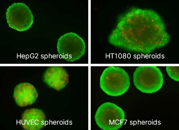
Spheroids of different cell lines were generated by the liquid overlay method and transferred to the Bioinert surface for FDA/PI staining and live cell imaging. FDA (green) indicates living cells at the outer spheroid layers. PI (red) marks dead cells within the necrotic spheroid center. Note the different spheroid morphology, which depends on the cell type. Microscope: Nikon Eclipse Ti, 4x objective lens.
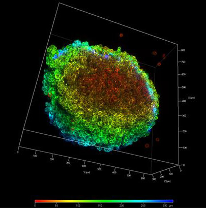
底部厚度符合 #1.5 蓋玻片,影像清晰精確

High resolution confocal microscopy on the Bioinert surface. Confocal laser scanning microscopy projection of an HT-1080 LifeAct spheroid. The colors indicate the distance from the surface. Warm colors = close to the surface, cold colors = distant from the surface.
特殊上蓋與皿身卡榫設計,可減少培養液蒸散,長時間細胞培養也 OK

產品規格
| Ø µ-Dish | 35 mm |
| Height without lid | 12 mm |
| Volume | 2 ml |
| Growth area | 3.5 cm² |
| Ø observation area | 21 mm |
| Bottom | ibidi Polymer Coverslip with Bioinert surface |
| Bioinert Surface thickness | 200 nm |
| Bioinert Surface material | Polyol-based hydrogel |
| Protein coatings | Not possible |
訂購資訊
| Product Name | Surface | Pack Size | Cat. No. |
|---|---|---|---|
| µ-Dish 35 mm, high Bioinert | Bioinert: #1.5 polymer coverslip, surface passivation with Bioinert, sterilized | 30 | IB-81150 |


