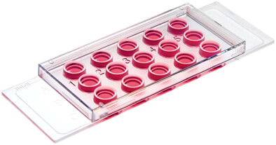
µ-Slide Angiogenesis

- 適用於血管新生 (tube formation assays)、血管萌芽 (sprouting assays)、3D 細胞培養 (3D cell culture)、活細胞影像觀察攝影 (live cell imaging)、細胞免疫螢光染色 (immunofluorescence) …等實驗。
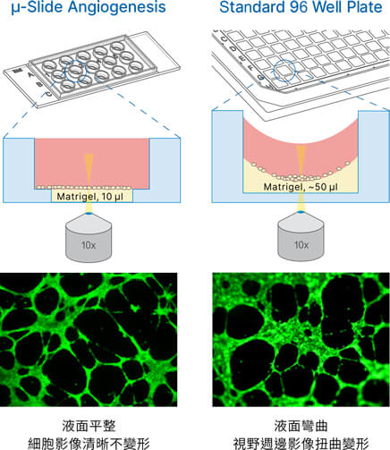
- 每個獨立 well 僅需 10 µl Matrigel® 即可進行實驗,與一般標準 96 孔盤相比可大幅節省 90% 的膠體用量!
- 可避免表面張力造成細胞影像扭曲變形的現象,有效提升細胞影像品質。
- 所有細胞皆可落於同一焦距上,方便影像觀察。
- Matrigel®, Collagen, Agarose... 等膠體皆可適用。
歡迎與我們聯繫索取更多 µ-Slide Angiogenesis 產品資訊與文獻。
產品特點
獨特「well-in-a-well」設計可大幅減少每次血管新生實驗所需的細胞、膠體、以及藥物用量,更可提供完美的影像品質

上圖左:「well-in-a-well」設計是指在 µ-Slide Angiogenesis 的每個獨立培養槽內 (直徑 5 mm),還含有一個 4 mm 直徑的槽體可供注入 gel matrix (例如 Matrigel®, collagen) 使用。此內槽僅需 10 µl 的 gel matrix 即可剛好注滿,形成一厚度達 0.8 mm 的平整細胞基質膠層。上圖右:注入在傳統細胞培養盤中的 gel matrix 往往會因為表面張力形成“圓弧形”的膠層,容易導致細胞生長分佈不均的情形,使得可觀察範圍縮小至槽體中央附近;而同樣的表面張力影響性,亦會造成膠層上方所灌注的培養液形成弧形液面,使得所觀察的細胞影像產生扭曲變形的現象。μ-Slide Angiogenesis 玻片的「well-in-a-well」設計完美解決了以上這兩大問題,使得影像可以更清晰且容易觀察,同時大幅減少 gel matrix 的使用量。
底部厚度符合 #1.5 蓋玻片,影像清晰易觀測

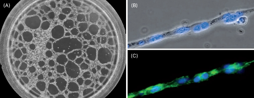
(A) Phase contrast image showing one well of the µ-Slide Angiogenesis with HUVEC cells on Matrigel® after 12 hours of incubation during a tube formation assay. Phase contrast (B) and fluorescence microscopy (C) of a single strand composed of HUVEC cells during a tube formation assay in the µ-Slide Angiogenesis. The F-actin cytoskeleton is stained green and the cell nuclei are stained blue.
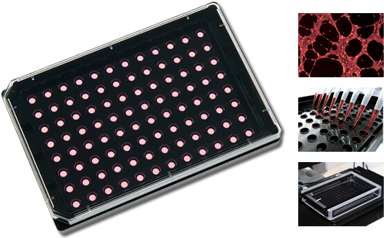
µ-Plate Angiogenesis 96 Well 具備 µ-Slide Angiogenesis 的一切特質與優點,應用於高通量 (high throughput) 實驗更划算便利

產品規格
µ-Slide Angiogenesis 與 µ-Plate Angiogenesis 96 Well 規格比較
| µ-Slide Angiogenesis |
µ-Slide Angiogenesis Glass Bottom |
µ-Plate Angiogenesis 96 Well |
|
|---|---|---|---|
| Outer dimensions | W25.5 x L75.5 mm | W25.5 x L75.5 mm | W85.5 x L127.7 mm |
| Number of wells | 15 | 15 | 96 |
| Volume inner well | 10 µl | 10 µl | 10 µ |
| Ø inner well | 4 mm | 4 mm | 4 mm |
| Depth inner well | 0.8 mm | 0.8 mm | 0.8 mm |
| Volume upper well | 50 µl | 50 µl | 70 µl |
| Ø upper well | 5 mm | 5 mm | 5 mm |
| Growth area inner well | 0.125 cm² | 0.125 cm² | 0.125 cm² |
| Coating area using 10 µl | 0.23 cm² | 0.23 cm² | 0.23 cm² |
| Bottom | ibidi Polymer Coverslip | Glass Coverslip No. 1.5H | ibidi Polymer Coverslip |
底部材質規格比較
| #1.5 ibidi Polymer Coverslip (ibidi 特殊塑料) |
#1.5H ibidi Glass Coverslip (#1.5H 高精度玻璃) |
|
|---|---|---|
| Bottom thickness | 180 µm (+10/-5 µm) | 170 µm (+/-5 µm) |
| Bottom material | Polymer | D 263 M Schott high precision glass |
| Gas permeability | Yes | No |
| Compatibility with protein coatings | Yes | Yes |
| Immersion oil compatibility | Yes | Yes |
| Autofluorescence | Low | Low |
| Transmission | Very high (even ultraviolet) | High (ultraviolet restrictions) |
| Refractive index (nD 589 nm) | 1.52 | 1.52 |
| Brightfield Microscopy | ++ | ++ |
| Phase Contrast | ++ | ++ |
| Differential Interference Contrast (DIC) | ++ | ++ |
| Widefield Fluorescence | ++ | ++ |
| Confocal Fluorescence | ++ | ++ |
| Total Internal Reflection Fluorescence (TIRF) | + | ++ |
| Super-Resolution Microscopy | + | ++ |
※更多材質特性比較資訊請見這裡。
訂購資訊
| Product Name | Surface | Pack Size | Cat. No. |
|---|---|---|---|
| µ-Slide Angiogenesis | ibiTreat: #1.5 polymer coverslip, tissue culture treated, sterilized | 15 | IB-81506 |
| Uncoated: #1.5 polymer coverslip, hydrophobic, sterilized | 15 | IB-81501 | |
| µ-Slide Angiogenesis Glass Bottom | Glass Bottom: #1.5H (170 µm +/- 5 µm) D 263 M Schott glass, sterilized | 15 | IB-81507 |
| µ-Plate Angiogenesis 96 Well | ibiTreat: #1.5 polymer coverslip, tissue culture treated, sterilized | 15 | IB-89646 |


