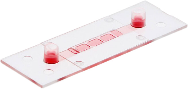
µ-Slide I Luer 3D

- 單一微通道含 3 個培養槽,可供注入 gel matrix (例如 Matrigel®, collagen) 形成一平整細胞基質膠層,搭配 ibidi Pump 使用即可進行流體環境 3D 細胞培養實驗 (3D cell culture under flow)。
- 上蓋與底部厚度皆符合 #1.5 蓋玻片標準,可直接結合高倍率光學或螢光顯微鏡進行細胞影像觀察。
- 產品應用包含:流體環境 3D 細胞培養、細胞極化試驗 (apical-basal cell polarity assays)、肺泡細胞或皮膚細胞微環境模擬、血腦障壁 (blood brain barrier) 模擬…等。
歡迎與我們聯繫索取更多 µ-Slide I Luer 3D 產品資訊與文獻。
產品特點
µ-Slide I Luer 3D 可搭配 ibidi Pump 使用,於流體環境進行 3D 細胞培養實驗


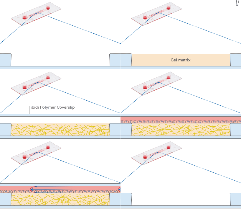
The μ-Slide I Luer 3D has three wells with one channel on top for culturing cells on a 3D gel matrix with defined flow. Each well can be filled with a gel, on which cells can be cultivated and microscopically investigated. The channel can be connected to ibidi Pump System for the application of defined shear stress. Using this method, an in vivo-like functional monolayer (e.g., an endothelial barrier) can be established on the gel matrix without any artificial filters or membranes involved. The μ-Slide I Luer 3D is especially designed for the simulation of blood vessels using endothelial cells.
µ-Slide I Luer 3D 應用彈性多元

The µ-Slide I Luer 3D is a versatile channel slide for a huge variety of cell culture applications using a 3D gel matrix and shear stress: (i) Establishing a cell monolayer on the gel matrix—investigate potential polarization effects, (ii) Culturing cells or cell clusters inside the gel matrix, and (iii) Performing assays using different gel matrices with varying concentration, stiffness, or chemical compounds. If cells are embedded inside the gel matrix, the perfusion delivers oxygen and nutrients to them.
µ-Slide I Luer 3D 上蓋與底部厚度皆符合 #1.5 蓋玻片,影像清晰易觀測

Phase contrast microscopy of HUVEC after culturing them under flow at 10 dyn/cm² for 2 days (A) and 5 days (B) on a Collagen Type I rat tail (2 mg/ml). Note the cobblestone-like cell morphology after 5 days of culture under flow. 20x objective. (C) Fluorescence microscopy of HUVEC after culturing them under flow at 10 dyn/cm² for 5 days on a Collagen Type I rat tail (2 mg/ml). Immunostaining of alpha-tubulin (red); the F-actin cytoskeleton was stained using phalloidin (green). Nuclei are stained with DAPI (blue). 10x objective.
產品規格
| Outer dimensions | W25.5 x L75.5 mm |
| Adapters | Female Luer |
| Number of wells | 3 |
| Volume of wells | 16 µl |
| Well dimensions | 5.4 x 4.0 mm |
| Well height (without channel) | 0.8 mm |
| Growth area per well | 0.21 cm² |
| Coating area per well | 0.34 cm² |
| Channel width | 5.0 mm |
| Channel height (without well) | 0.6 mm |
| Channel volume (without wells) | 150 μl |
| Volume per reservoir | 60 μl |
| Top cover | ibidi Polymer Coverslip |
| Bottom | ibidi Polymer Coverslip |
訂購資訊
| Product Name | Surface | Pack Size | Cat. No. |
|---|---|---|---|
| µ-Slide I Luer 3D | ibiTreat: #1.5 polymer coverslip, tissue culture treated, sterilized | 15 | IB-87176 |
| Uncoated: #1.5 polymer coverslip, hydrophobic, sterilized | 15 | IB-87171 |
流體環境 3D 細胞培養產品比較表
 µ-Slide Spheroid Perfusion |
 µ-Slide III 3D Perfusion |
 µ-Slide I Luer 3D |
|
|---|---|---|---|
| Perfusion of samples | ✓ | ✓ | ✓ |
| Defined shear stress on cell monolayers | ✕ | ✕ | ✓ (on gel matrix) |
| Gel matrices for 3D | ✕ | ✓ | ✓ |
| Spheroids/organoids | free floating in well | inside gel matrix only | inside gel matrix only |
| Suspension cells | free floating in well | inside gel matrix only | inside gel matrix only |
| Adherent cells | on coverslip | inside or on gel matrix | inside or on gel matrix |


